Formalin fixation is a critical step in the preservation of biological tissues. As many who read this are likely laboratory professionals, you may already be familiar with this process. Here’s a brief explanation of how it works step by step:
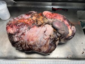
- Collection of Tissue Sample: The process typically begins when a tissue sample, such as a biopsy or surgical specimen, is obtained from a patient during a medical procedure.
- Transport to the Laboratory: The tissue sample is then carefully transported to the pathology laboratory. It’s crucial to minimize any delays between the collection and fixation to ensure the integrity of the specimen.
- Preparation for Fixation: In the laboratory, the tissue sample is prepared for fixation. This often involves trimming and sizing the tissue to a manageable and uniform thickness, which facilitates the penetration of fixative.
- Immersion in Formalin: Formalin, a solution of formaldehyde gas dissolved in water, is the most commonly used fixative. The tissue is completely immersed in a container filled with formalin. The ratio of formalin to tissue volume should be sufficient to ensure thorough fixation.
- Fixation Duration: The duration of fixation varies depending on the size and type of tissue. Typically, small biopsies may require 6-24 hours, while larger tissue specimens from surgeries may need several days or even weeks of fixation.
- Fixation Penetration: During the fixation process, formalin penetrates the tissue and reacts with proteins, forming cross-links. This chemical reaction stabilizes cellular structures and prevents enzymatic degradation, essentially “freezing” the tissue in its current state.
- Tissue Storage: After the appropriate fixation time has elapsed, the tissue is removed from the formalin solution. Excess formalin is drained, and the tissue is often stored in a different solution, such as 70% ethanol, until further processing, such as embedding in paraffin wax or preparation for histological examination.
- Histological Processing: Once fixed, tissues can undergo additional processing steps, including dehydration, clearing, and embedding in paraffin wax. These steps prepare the tissue for thin sectioning, which is essential for microscopic examination.
- Microscopic Analysis: The fixed and processed tissue sections are cut into thin slices, placed on glass slides, and stained with various dyes to highlight specific structures and cell types. Pathologists then examine these slides under a microscope to make diagnostic assessments.
Formalin fixation is crucial in preserving tissue integrity and cellular structures, allowing pathologists to study and diagnose various diseases. Your work in the path lab will likely involve ensuring that this process is carried out accurately to provide high-quality diagnostic information to clinicians and researchers


