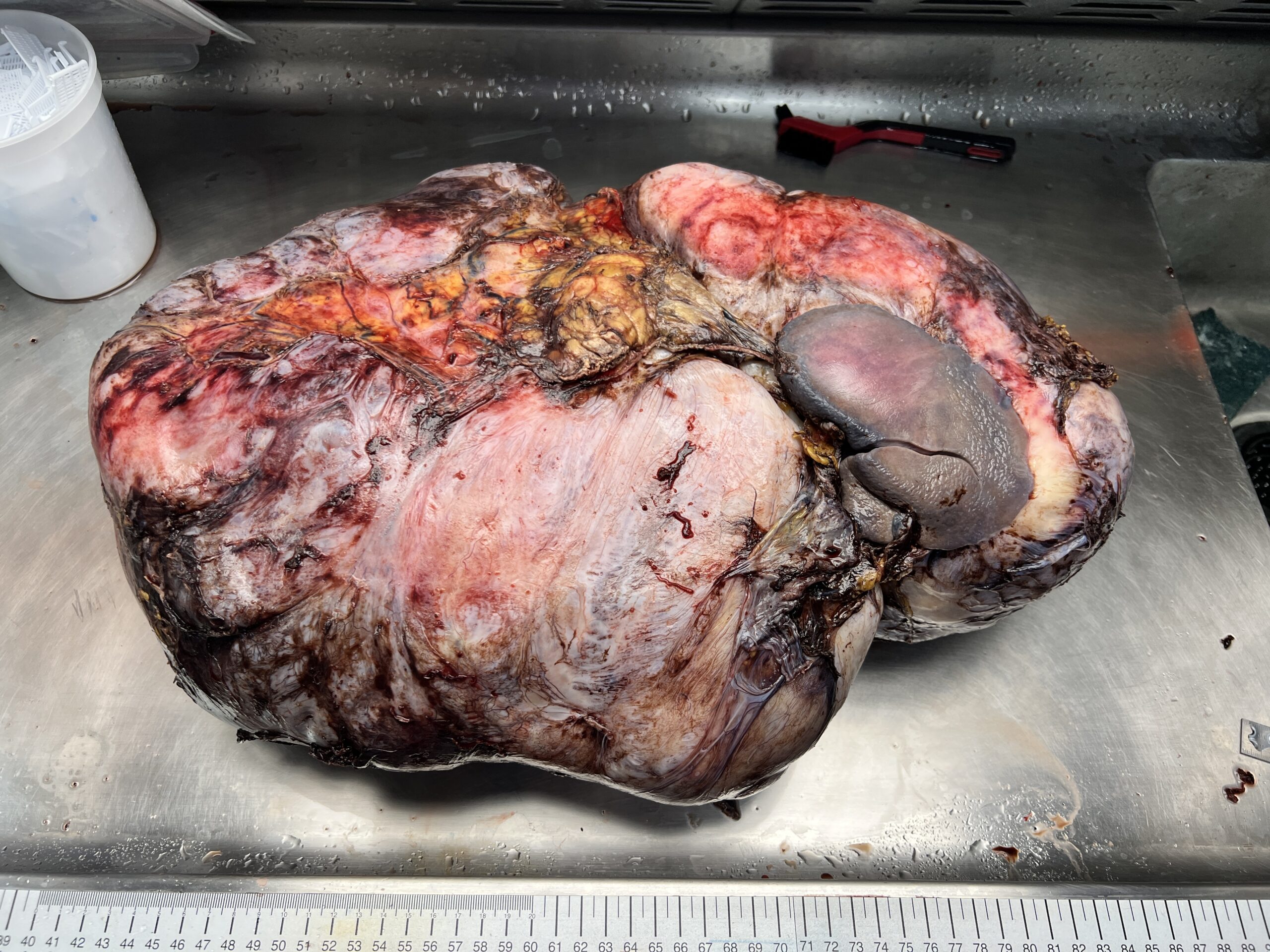
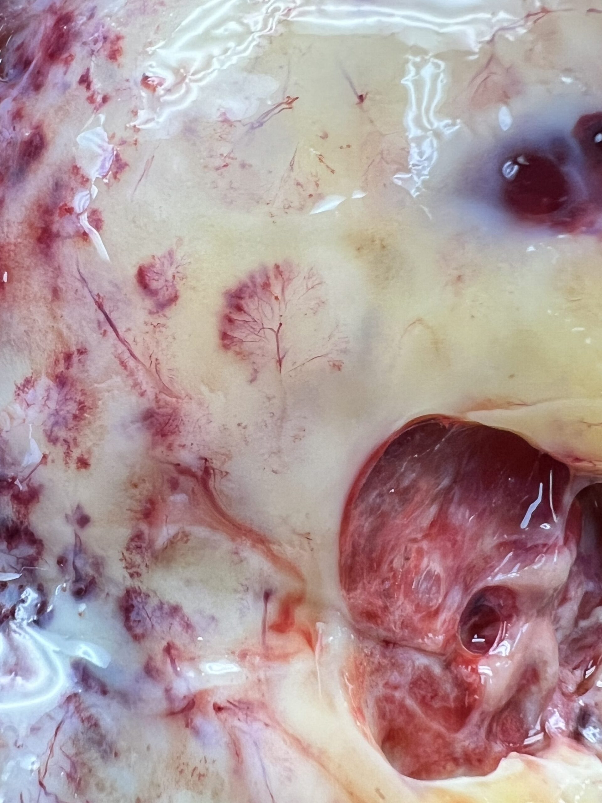
This large mass was located in the upper abdomen. It had involved the spleen, pancreas and stomach. This made it impossible for the surgeon to remove the mass without also removing all or part of each of those organs. Most of the specimen consisted of a somewhat gelatinous, white-tan material, which I could see as I sectioned through the it with the largest blade I had available. The gelatinous nature of the tissue made it so I could fairly clearly make out the small blood vessels running throughout the whole thing, which looked super cool.
My job in examining this specimen was to determine just how involved the mass had become in relation to the attached organs so I had to slice through the spleen, pancreas and stomach to see if they were just connected or if the tumor had started to eat away at them.
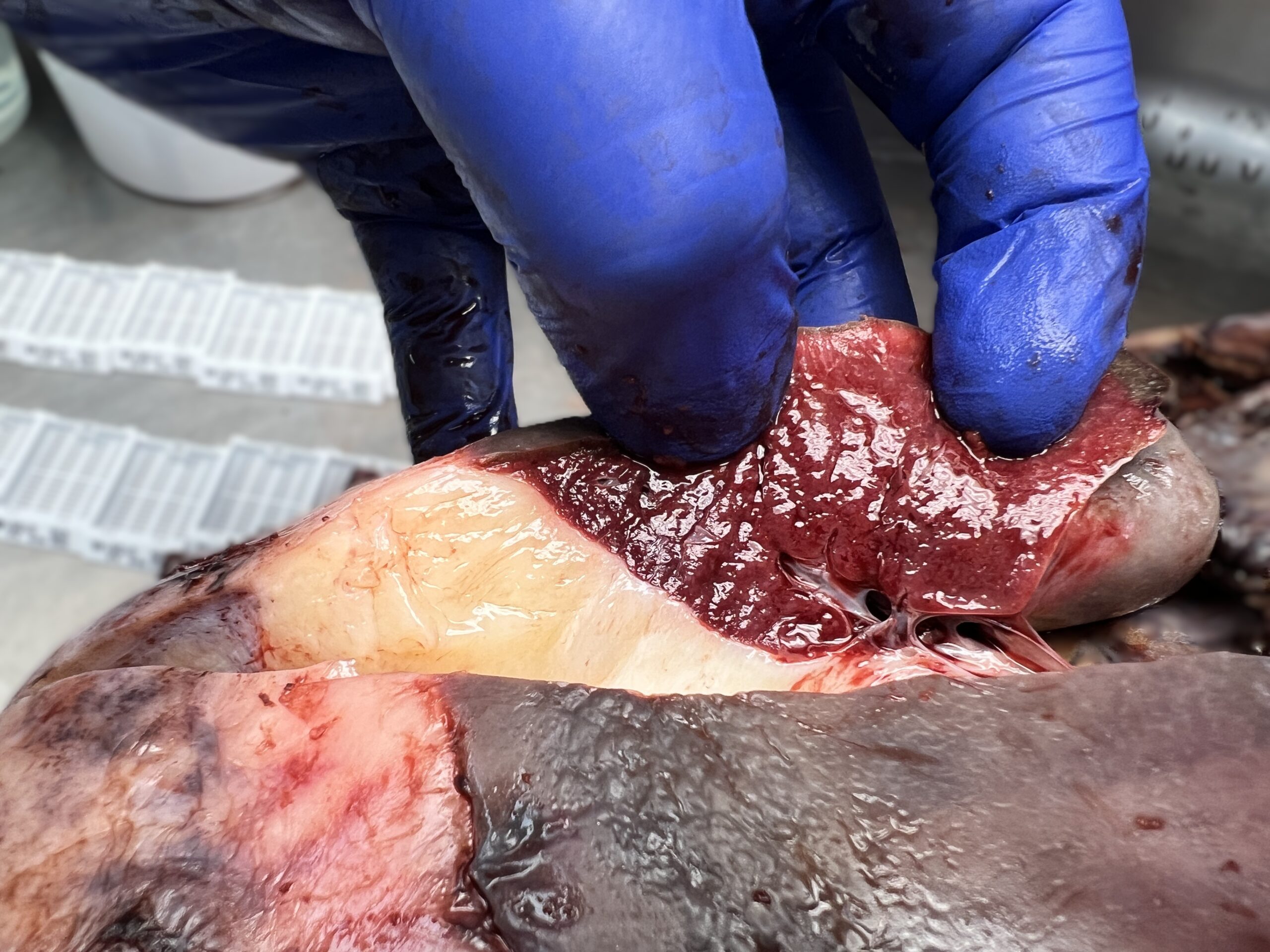
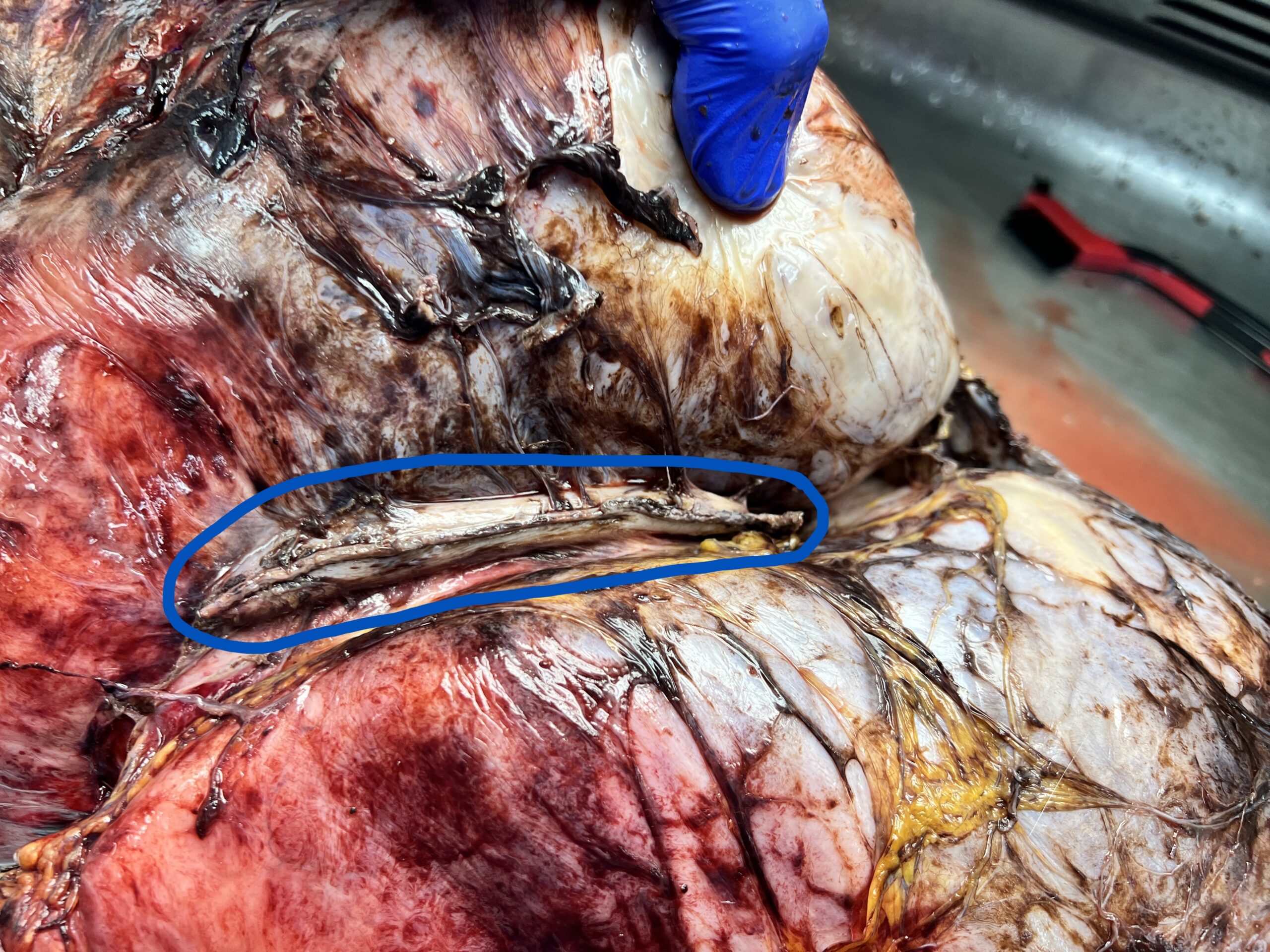
When I cut into the spleen I was surprised to see the stark contrast between the two different tissue types. It was clear there was no connective tissue between the two but I wasn’t able to see with the naked eye whether or not there was any overlap. Usually when a tumor invades into another organ the line isn’t so straight or well-defined.
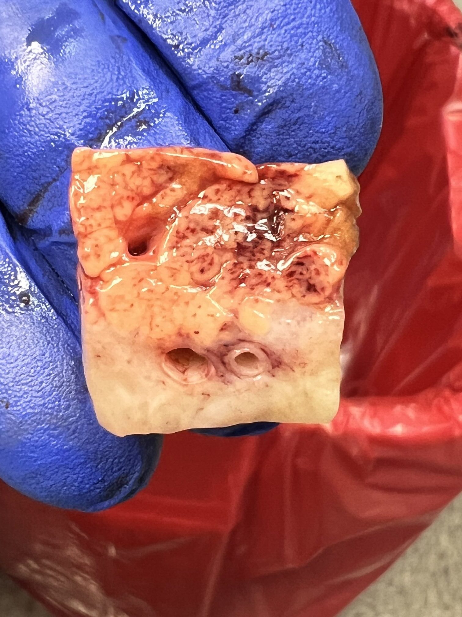
However, as I cut into the pancreas it was much less clear where the tumor stopped and the pancreas started. This is usually a more clear sign of invasion. Sometimes the pancreas and the surrounding fat can have a similar appearance, as was the case here.
The stomach was not involved by the tumor at all. However, the outer surface was covered in dense fibrous adhesions which make it impossible for a surgeon to tell. So part of the stomach had to be removed as a precaution.
Final diagnosis: Desmoid type fibromatosis with invasion of the pancreas
Overall this specimen was fascinating and I really enjoyed exploring all the unique complications it brought!